

92 F. D. Fragiskos
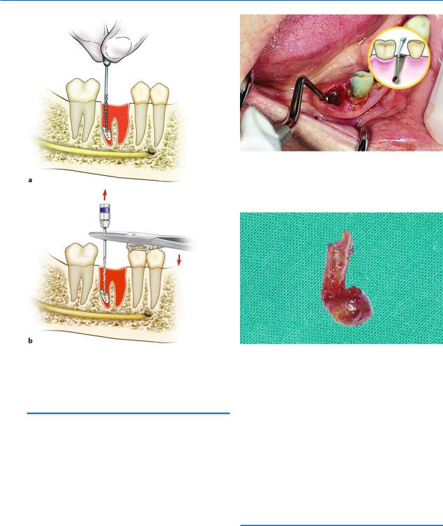
Fig. 5.42 a, b. Removal of the root tip using an endodontic file. After the endodontic file enters the root canal, the root tip is drawn upwards by hand (a), or with a needle holder (b)
Fig. 5.43. Curettage of the socket after tooth extraction for removal of the periapical lesion
Fig. 5.44. Root, together with firmly attached lesion at tip of root, right after extraction
5.6
Postextraction Care of Tooth Socket
After extraction of the tooth, the bottom of the socket is curetted (as long as the tooth is nonvital) with a periapical curette, to remove any periapical lesion from the area (Fig. 5.43). Curetting must be done carefully, because if any remnants of granulation tissue remain in the socket, there is a chance they will develop into a cyst, because a large percentage contain epithelial cells. Sometimes the lesion is firmly attached to the root tip of the tooth and is extracted together with the tooth (Fig. 5.44). Even in this case, the socket must be inspected, but only in the apical region. When the lesion is large and the entire lesion cannot be removed through the socket alone, then surgery is required.
Afterwards, and only if considered necessary (e.g., there are sharp bone edges), the alveolar margin is smoothed using rongeur forceps or a bone file, and then the lingual and buccal plates are compressed using finger pressure. This is done to restore the expansion of the socket caused by the extraction, and also for initial control of hemorrhage. Hemostasis is also aided by the patient applying pressure on gauze placed over the socket for 30–45 min.
5.7
Postoperative Instructions
After finishing the surgical procedure, oral and written instructions are given to the patient, concerning exactly what to do in the next few days. These instructions normally include the following:

Chapter 5 Simple Tooth Extraction |
93 |
ΟRest: After surgery, the patient should stay at home and not go to work for 1 or 2 days, depending on the extent of the surgical wound and the patient’s physical condition.
ΟAnalgesia: Take a painkiller (but not salicylates, aspirin), every 4 h, for as long as the pain persists.
ΟEdema: After the surgical procedure, the extraoral placement of cold compresses (ice pack wrapped in a towel) over the surgical area is recommended.
This should last for 10–15 min at a time, and be repeated every half hour, for at least 4–6 h.
ΟBleeding: The patient must bite firmly on gauze placed over the wound for 30–45 min. In case bleeding continues, another gauze is placed over the wound for a further hour.
ΟAntibiotics: These are prescribed only if the patient has certain medical conditions or inflammation (see Chaps. 1 and 16).
ΟDiet: The patient’s diet on the day of the surgical procedure must consist of cold, liquid foods (pudding, yogurt, milk, cold soup, orange juice, etc.).
ΟOral hygiene: Rinsing the mouth is not allowed for the first 24 h. After this, the mouth may be rinsed with warm chamomile or salt water, three times a day for 3–4 days. The teeth should be brushed with a toothbrush and flossed, but the patient should avoid the area of surgery.
ΟRemoval of sutures: If sutures were placed on the wound, the patient must have them removed a week later.
Bibliography
Archer WH (1975) Oral and maxillofacial surgery, 5th edn. Saunders, Philadelphia, Pa.
Bean LR, King DR (1971) Pericoronitis: its nature and etiology. J Am Dent Assoc 83:1074–1077
Byrd DL (1971) Exodontia: modern concepts. Dent Clin North Am 15:273–298
Cerny R (1978) Removing broken roots: a simple method. Aust Dent J 23:351–352
Gans BJ (1972) Atlas of oral surgery. Mosby, St. Louis, Mo. Guralnick WC (1968) Textbook of oral surgery. Little Brown,
Boston, Mass.
Hayward JR (1976) Oral surgery. Thomas, Springfield, Ill. Hooley JR, Whitacre RA (1986) A self-instructional guide
to oral surgery in general practice, vol 2, 5th edn. Stoma, Seattle, Wash.
Howe GL (1996) The extraction of teeth, 2nd edn. Wright, Oxford
Howe GL (1997) Minor oral surgery, 3rd edn. Wright, Oxford
Javaheri DS, Garibaldi JA (1997) Forceps extraction of teeth with severe internal root resorption. J Am Dent Assoc 128(6):751–754
Kamberos S, Kolokoudias M, Stavrou E, Vagenas N, Fragiskos F (1989) Frequency and causes of extraction of permanent teeth. A ten-year (1968–1977) clinicostatistical investigation. Odontostomatologike Proodos 43:423– 433
Killey HC, Seward GR, Kay LW (1975) An outline of oral surgery. Part I. Wright, Bristol
Koerner KR (1995) Predictable exodontia in general practice. Dent Today 14(10):52, 54, 56–61
Koerner KR, Tilt LV, Johnson KR (1994) Color atlas of minor oral surgery. Mosby-Wolfe, London
Kruger E (1979) Oral surgery. Laterre, Athens
Kruger GO (1984) Oral and maxillofacial surgery, 6th edn. Mosby, St. Louis, Mo.
Laskin DM (1985) Oral and maxillofacial surgery, vol 2. Mosby, St. Louis, Mo.
Lehtinen R, Ojala T (1980) Rocking and twisting moments in extraction of teeth in the upper jaw. Int J Oral Surg 9(5):377–382
Leonard MS (1992) Removing third molars: a review for the general practitioner. J Am Dent Assoc 123:77–86
Lopes V, Mumenya R, Feinmann C, Harris M (1995) Third molar surgery: an audit of the indications for surgery, post-operative complaints and patient satisfaction. Br J Oral Maxillofac Surg 33:33–35
Malden N (2001) Surgical forceps techniques. Dent Update 28(1):41–44
Martis CS (1990) Oral and maxillofacial surgery, vol 1. Athens
Masoulas G (1984) Tooth extractions. Athens
Ojala T (1980) Rocking moments in extraction of teeth in the lower jaw. Int J Oral Surg 9(5):367–372
Papadopoulos AD (1976) Tooth extractions. Athens Peterson LJ, Ellis E III, Hupp JR, Tucker MR (1993) Contem-
porary oral and maxillofacial surgery, 2nd edn. Mosby, St. Louis, Mo.
Quinn JH (1997) The use of rotational movements to remove mandibular molars. J Am Dent Assoc 128(12):1705– 1706
Rounds CE (1962) Principles and technique of exodontia, 2nd edn. Mosby, St. Louis, Mo.
Sailer HF, Pajarola GF (1999) Oral surgery for the general dentist. Thieme, Stuttgart
Stefanopoulou-Vrettou V (1966) Practical oral surgery (tooth extractions). Athens
Sullivan SM (1999) The principles of uncomplicated exodontia: simple steps for safe extractions. Compendium Cont Educ Dent 20(3 Spec No):3–9, quiz 19
Thoma KH (1969) Oral surgery, vol 1, 5th edn. Mosby, St. Louis, Mo.
Waite DE (1987) Textbook of practical oral and maxillofacial surgery, 3rd edn. Lea and Febiger, Philadelphia, Pa.
Zambito RF, Zambito ML (1992) Exodonture. Technique and art. NY State Dent J 58(3):33–37